Welcome to
the University of Wisconsin-Madison
Department of Geoscience
Eugene Cameron EPMA Lab
Soft X-Ray EPMA Research
Update July 21, 2019
Hot of the press! June 2019 QMA poster showing some examples of our low kV EPMA
Traditionally, EPMA has been done at 15-20 keV, for a variety of reasons, principal ones being:
- Utilization of the Ka lines of the transition metals (not accessible at lower keVs), which are better characterized for matrix correction,
- And in many--not all--cases do not have peak shifts associated with bonding differences between standards and unknowns)
- And these voltages tend to minimize surface effects (roughness, oxide skins, discrepancies in conductive coating thicknesses, etc) which add to the imprecise and inaccuracy of the analyses
For the most part, the electron source was the hair-pin W filament, whose spot size on the sample, at typical probe currents (20-30 nA) would be in the 0.5-1 micron diameter range. So the smallest typical 'normal' (eye of the beholder) object (say a feldspar crystal) might be in the 2-3 micron diameter range. Forget about a 1 um exsolution lamella in a pyroxene or ilmenite.
With the emergence of the Field Emission Electron Probe (JEOL in 2003 and CAMECA in 2011) and the ability to shrink the electron beam diameter by a factor of 5-10, it has now become possible to consider EPMA of sub-micron size features--IF BEAM ENERGY (KV) IS DROPPED. This depends upon several factors, a combination of (1) the beam diameter (itself a combination of the keV and nA being used), (2) the incident electrons' scattering distance (lateral and depth) in the specimen, and (3) the edge energy required to excite a particular X-ray line.
Starting in 2011, we began investigating low voltage (5 keV) EPMA of iron silicides, resulting from the discovery by UW Madison researchers of micron and sub-micron iron silicides in an impacted lunar feldspar grain. Phil Gopon's Ph.D. research focuses upon both this lunar iron silicide as well as the application of low voltage EPMA to terrestrial samples. Phil's lunar iron silicide EPMA research has now been published (2013) in Microscopy and Microanalysis. He reports on the benefit of utilizing an otherwise ignored iron L X-ray line, the LL line, which is not involved with chemical bonding AND is far away from the L3 edge (the nemesis of the La X-ray line), both which make use of the Fe La line next to impossible for EPMA. (Fe Lb is not much better).
In August 2016, Aurélien Moy (PhD, Montpelier with Claude Merlet) began an NSF-funded post doc to continue this research. One main goal was to go back to the iron La/Lb X-ray lines, and see if it was possible to "crack the code". While the Fe LL line is an easy solution, this line is of less value in many minerals and glasses where the iron content is not a high as in the binary iron silicides. After much effort to decipher the issue of mass absorption coefficients, Moy went back to the theoretical underpinnings of the spectra collection in EPMA and found that the combination of the imprecision of the WDS spectrometers AND the natural peak broadening in the context of the absorption edge being in the middle of it all, made the MAC approach impossible for this situation. Instead, an old empirical approach was found to be a solution: using standards to generate "Fe La/lb peak integral" K-ratios plotted against Fe content, an equation was generated and could be applied to unknown samples. This was first published in mid 2019 in Microscopy and Microanalysis for iron silicides, and soon after in American Mineralogists for olivines. Work continues on other Fe-bearing minerals.
Other low voltage research projects which we have been involved with:
- Low voltage EPMA of stainless steel. Xavier Llovet organized a round robin of many labs to attempt low voltage EPMA of this common industrial material. The results (using La lines) indicated there are many problems which need to be resolved. This was published in 2012.
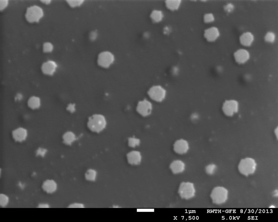
- K409 is an interesting specimen, a glass with ~800 nm crystals of magnetite, of utility in evaluating electron beam diameters (e.g., FE, LaB6, W guns) and also the ability to estimate the X-ray analytical resolution for the elements present in the glass and the crystal. It was produced by NBS/NIST 30-40 years ago and because it was heterogeneous, was never distributed. We have some of this material available for distribution. Cathey, Gopon and Fournelle (2013) discuss it at the December 2013 AGU conference. Contact John Fournelle if you would like some of this material (~1 mm chip): johnf at geology dot wisc dot edu
A 2012 paper by Merlet and Llovet with a formulation for x-ray analytical resolution that combines both the electron beam diameter and the x-ray range (lateral and depth)
Most of the low voltage EPMA work going on is happening in Europe, and the 2014 EMAS (European Microbeam Analysis Society) conference in Porto, Portugal, features many talks and posters dealing with low voltage EPMA, FE EPMA and critical issues such as carbon contamination.
We thank and acknowledge our funder (NSF-EAR) and colleagues who have assisted our ongoing low voltage EPMA research, including Xavier Llovet (U of Barcelona) and Peter Sobol (UW-Madison), and particularly those in making use of their Field Emission microprobes available to us: Bill Horn at Exxon-Mobil (JXA-8530), David Snoeybous, Michel Outrequin, Chrystel Hombourger (CAMECA, SXFive-FE), Silvia Richter and Philippe Pinard (RWTH Aachen, JXA-8530) and Henny Cathey (ASU, JXA-8530, now at Brisbane QUT).
Some older publications references low voltage x-ray lines (e.g. M lines of REE)
For more information, contact:
John Fournelle
Dept of Geology and Geophysics
University of Wisconsin-Madison
1215 W. Dayton St
Madison, WI 53706
(608) 262-7964 (office) 265-4798 (lab) 262-0693 (fax)
johnf*at*geology dot wisc dot edu
Go to Electron Microprobe Lab page